Retinal Imaging
Standard fundus imaging captures the optic nerve, macula and the first branches of the retinal vasculature.
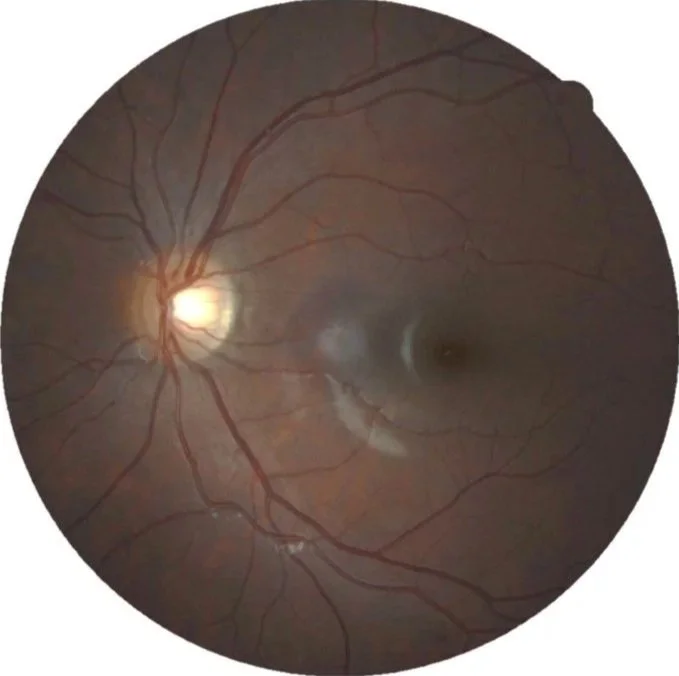
Canon CR-1 standard fundus imaging
Fundus Photography
Zeiss Clarus 500
Heidelberg Spectralis
Canon CR-1
Widefield and ultra-widefield fundus imaging captures the central and far peripheral retinal tissue, capturing data about the retina in its entirety. It also uses similar technology to the MultiColor, where red, green and blue reflectance highlight different retinal structures.

Clarus widefield fundus imaging

Clarus ultra-widefield fundus imaging with three different wavelengths of light and true colour
MultiColor fundus imaging captures all of the same tissues as standard fundus imaging, but uses three different laser wavelengths to better visualise different retinal layers. Blue reflectance highlights highlights the retinal nerve fibres (important for optic nerve pathology like glaucoma, as well as traction pathology like epiretinal membranes). Green reflectance highlights the retinal vasculature (important for detecting abnormal blood flow, especially in conditions like diabetes and hypertension). Infrared reflectance highlights the last layers of the retina and its underlying blood supply (important for pathology like macular degeneration and retinal dystrophies). The MultiColor image compiles all three laser images into a single image.

Heidelberg Spectralis MultiColor imaging
Fundus Auto-fluorescence (FAF)
Zeiss Clarus 500
Blue autofluorescence uses light of a specific wavelength to stimulate retinal cells, which autofluoresce in response. Cells that are highly metabolically active fluoresce brightly while cells that are atrophic do not fluoresce at all.

Blue autofluorescence uses light of a specific wavelength to stimulate retinal cells, which autofluoresce in response. Cells that are highly metabolically active fluoresce brightly while cells that are atrophic do not fluoresce at all.
Optic nerve and macula scanning identifies all retinal layers (inner limiting membrane, retinal nerve fibre layer, ganglion cell layer, inner plexiform layer, inner nuclear layer, outer plexiform layer, outer nuclear layer, external limiting membrane, photoreceptors, retinal pigment epithelium, Bruch’s membrane as well as choroid) in raster scans and quantifies thickness in printed reports. An ophthalmic professional can also analyse for abnormalities in optic nerve and macular architecture not detected by thickness reports. Changes from baseline scans are shown in progression displays.
Heidelberg Spectralis
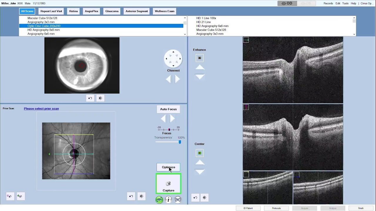
Posterior OCT (Optical Coherence Tomography) & Angiography


OCT angiography can also be used to assess blood flow at the macula and optic nerve.

Much like the Heidelberg Spectralis, the Zeiss Cirrus captures macular and optic nerve raster scans, thickness maps, progression displays and OCT angiography.
Zeiss Cirrus 6000 Angioplex
Posterior OCT (Optical Coherence Tomography) & Angiography