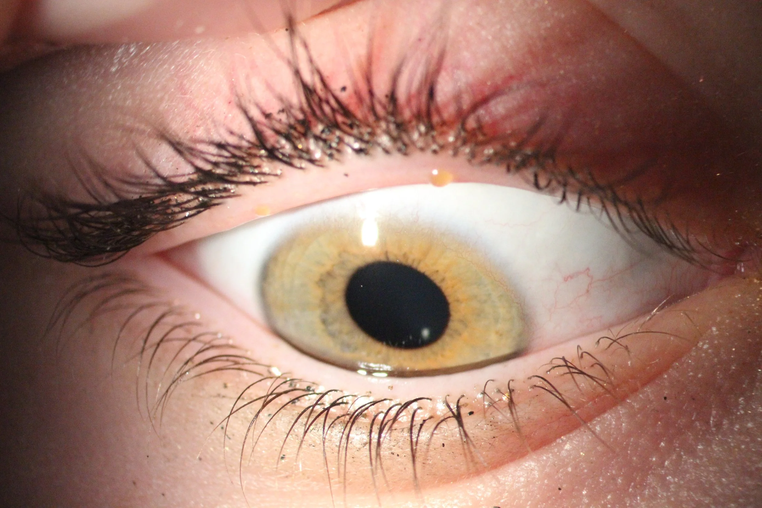
Anterior Eye Imaging
Slit lamps equipped with photography instrumentation are able to image the front of the eye, including the eyelids, conjunctiva, sclera, cornea, iris, anterior chamber, lens and anterior vitreous.
Zeiss SL800
Slit Lamp Biomicroscopy

Equipped with Wratten filters and cobalt blue light, these slit lamps are also excellent for ocular surface assessment with vital stains including sodium fluorescein and lissamine green.
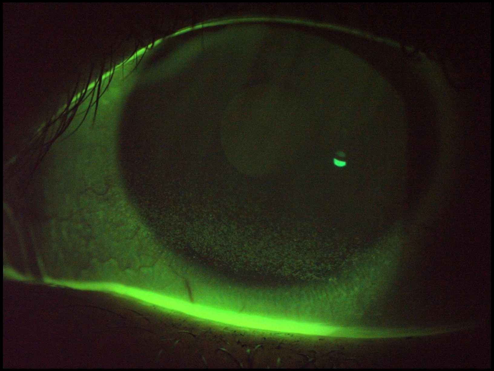
Much like the Zeiss slit lamp, the Takagi SM-70N is used to image the anterior eye and is also equipped with a Wratten filter and cobalt blue lighting.
Takagi SM-70N
Slit Lamp Biomicroscopy
This instrument uses reflective rings to capture an infrared image of the anterior cornea, through which corneal diameter (HVID) can be measured. The quality of the tear film can be analysed with these reflected rings. The scan provides quantitative data about the anterior cornea (Placido imaging) including:
Keratometry values
Corneal elevation
Refractive power
Tangential and axial maps
Shape descriptors such as e, p, Q and IS values
Two maps captured on the Medmont E300 can be compared using a difference map, which can be useful in showing change over time.
Medmont E300
Corneal Topography


Corneal tomography is captured using Schleimpflug imaging to provide data about the anterior and posterior cornea, anterior sclera, anterior chamber, anterior iris and anterior lens. This data includes:
Keratometry readings
Corneal thickness
Anterior and posterior corneal elevation
Corneoscleral height
Refractive power
Tangential and axial maps
Shape descriptors such as e, p, Q and IS values
Schleimpflug images (infrared photos and B-scans of the anterior eye)
Corneal diameter (HVID)
Ocular wavefront aberrometry
Lens retroillumination image
Oculus Pentacam Wave AXL
Corneal Tomography


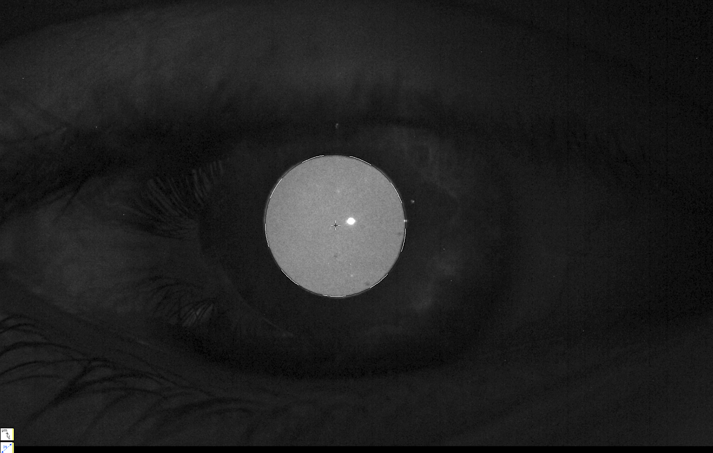
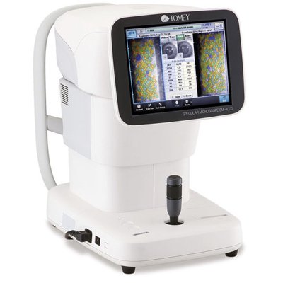
Specular microscopy is a non-invasive means of collecting information on the cornea and specifically the corneal endothelium at the central, midperipheral and peripheral cornea. The Tomey EM-4000 analyses up to 300 cells of the corneal endothelium over a 0.25x0.54mm capture area, measuring:
Cell density (CD)
Average cell area (AVG)
Coefficient of variation of cell area (CV)
Percentage of hexagonal cells (6A)
Tomey EM-4000
Specular Microscopy
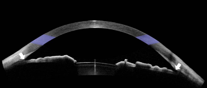
Anterior OCT captures information including corneal thickness (central and peripheral) and corneal transparency. It can also be focussed to give information about iris contour and anterior chamber angle patency.
Heidelberg Spectralis
Anterior OCT

Much like the Heidelberg Spectralis, this OCT captures corneal thickness and transparency data, iris contour and anterior chamber angle patency. However, it can also perform epithelial thickness mapping on the cornea.
Zeiss Cirrus 6000 Angioplex
Anterior OCT